Man has always donned the hat of an inventor in his struggle for existence. The desire to grow and understand the complexity of nature has often led to innovation and technological advancement, the roots of which are deeply embedded in our understanding of basic sciences. In late 1940’s Felix Bloch and Edward Purcell discovered Nuclear Magnetic Resonance (NMR) where the magnetic property of atomic nuclei gets manipulated by external magnetic field which then helps in analyzing molecules, determining structures of proteins etc. Though this technological breakthrough could now enable us to scrutinize the structure of compounds, imaging a tissue or an organism to get minute structural details was a far cry and a huge technological challenge.Paul C. Lauterbur and Peter Mansfield conducted landmark research that lead to discovery and development of Magnetic Resonance Imaging (MRI) - one of the most useful imaging modalities in medicine today and were awarded the Nobel Prize in physiology or medicine in 2003 for their seminal contributions in this field. Lauterbur very carefully introduced small variations in the strength of the magnetic field and demonstrated that these variations can distinguish hydrogen nuclei in different parts of a sample. He was the first to construct an image based on NMR signals by the application of magnetic field gradients in different directions through the sample.Peter Mansfield on the other hard worked very extensively to develop efficient ways by which one could acquire NMR signals and construct these images at greater speed.MRI has widespread applications in medical diagnostics/ research and since then has revolutionized healthcare, saved millions of lives and improved the quality of life of people all
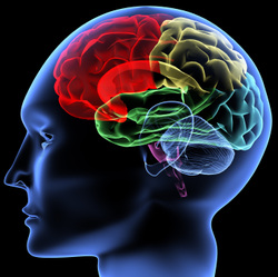
over the globe. MRI is an excellent noninvasive biophysical imaging technique used to probe almost all organs of body especially for the detailed and precise multi –directional cross sectional structural examination of the brain and the spinal cord and other soft tissues. MRI can also provide information about how the blood moves through certain organs and blood vessels. More than 60 million MRI scans have been performed worldwide since the first commercial MRI set up was made available around 1980’s.The method does not use ionizing radiation and thus has great advantage over X ray or computer tomography methods. Moreover since MRI provides detailed 3D images of tissue it can be extensively used to guide surgeons to identify lesions during complex neurosurgeries or to place electrodes during treatment of neurodegenerative disorders. MRI is also very extensively used in cancer diagnostics and accessing the extent of damage in accident victims. MRI has led to a paradigm shift in the world of disease diagnostics and treatment and thus finds widespread application all over the world.
over the globe. MRI is an excellent noninvasive biophysical imaging technique used to probe almost all organs of body especially for the detailed and precise multi –directional cross sectional structural examination of the brain and the spinal cord and other soft tissues. MRI can also provide information about how the blood moves through certain organs and blood vessels. More than 60 million MRI scans have been performed worldwide since the first commercial MRI set up was made available around 1980’s.The method does not use ionizing radiation and thus has great advantage over X ray or computer tomography methods. Moreover since MRI provides detailed 3D images of tissue it can be extensively used to guide surgeons to identify lesions during complex neurosurgeries or to place electrodes during treatment of neurodegenerative disorders. MRI is also very extensively used in cancer diagnostics and accessing the extent of damage in accident victims. MRI has led to a paradigm shift in the world of disease diagnostics and treatment and thus finds widespread application all over the world.


 RSS Feed
